human anatomy laboratory manual with cat dissections
This manual provides a comprehensive guide for exploring human anatomy through cat dissections, offering 30 exercises, full-color visuals, and practical applications for real-world learning.
Purpose and Benefits of the Manual
The Human Anatomy Laboratory Manual with Cat Dissections is designed to provide students with a hands-on, interactive learning experience, aligning lab activities with real-world clinical applications. Its purpose is to bridge the gap between theoretical knowledge and practical skills through 30 exercises covering all body systems. The manual benefits students by offering a clear, engaging writing style, full-color visuals, and tools like Visual Summary Tables and Why This Matters boxes. These features enhance understanding, retention, and the ability to apply anatomical concepts in healthcare settings. It supports diverse learning styles and prepares students for future careers in medicine and allied health fields.
Structure and Organization of the Manual
The manual is organized into 30 exercises, each focusing on specific body systems and anatomical structures. It begins with foundational concepts, such as anatomical terminology and the anatomical position, before progressing to detailed dissection activities. Exercises are supported by full-color illustrations, Visual Summary Tables, and Why This Matters boxes to enhance comprehension. The manual also includes pre-lab materials, such as video coaching, to prepare students for hands-on activities. Its logical flow and clear structure make it easy for students to follow, ensuring a systematic and engaging learning experience.

Key Features of the Manual
Engaging writing style, full-color illustrations, and 30 exercises covering all body systems. Includes Visual Summary Tables and Why This Matters boxes for enhanced learning and retention.
Engaging Writing Style and Full-Color Illustrations
The manual features a clear, engaging writing style that simplifies complex anatomical concepts, making them accessible and easy to understand. Full-color illustrations provide vivid, detailed visuals of anatomical structures, aiding students in identifying and understanding key features during dissections. These visuals are carefully designed to enhance learning and retention, ensuring students can visualize the relationships between different body systems.
The combination of an engaging narrative and high-quality images creates an immersive learning experience, helping students connect theoretical knowledge with practical observation. This approach fosters a deeper understanding of human anatomy through hands-on exploration and visual reinforcement.
Visual Summary Tables and Why This Matters Boxes
Visual Summary Tables condense complex anatomical information into organized, easy-to-review formats, helping students quickly compare and contrast structures across body systems. These tables highlight key features, functions, and relationships, making them invaluable for study and review. Accompanying Why This Matters boxes connect lab activities to real-world clinical scenarios, emphasizing the practical relevance of anatomical knowledge. This unique feature helps students appreciate how the concepts they learn apply to healthcare and everyday life, bridging the gap between theory and application.
Together, these tools enhance understanding, retention, and engagement, providing a comprehensive and meaningful learning experience tailored to future healthcare professionals.

Lab Exercises and Activities
The manual includes 30 comprehensive exercises covering all body systems, providing hands-on experience with cat dissections and other anatomical explorations to enhance practical understanding.
Hands-On Lab Experience with Cat Dissections
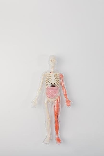
The manual offers a comprehensive hands-on lab experience, featuring 30 exercises that guide students through detailed cat dissections. Each exercise is designed to explore specific body systems, providing a practical understanding of anatomical structures. Full-color illustrations and clear instructions accompany each dissection, ensuring students can visualize and identify key components. The exercises also extend to organ and body dissections of other animals, such as sheep and cows, offering a comparative perspective. This interactive approach allows students to connect theoretical knowledge with real-world applications, enhancing their ability to understand human anatomy through direct observation and exploration.
Organ and Body Dissection Activities
The manual includes detailed organ and body dissection activities, emphasizing hands-on exploration of anatomical structures. Students engage in dissections of cats, as well as sheep and cows, to compare and contrast anatomical features. These exercises provide a comprehensive understanding of organ systems and their relationships. Clear instructions and full-color visuals guide students through each dissection, ensuring accurate identification of key structures. The activities are designed to align with human anatomy, helping students draw connections between animal models and human physiology. This practical approach enhances learning by combining observation with tactile experience, fostering a deeper appreciation of anatomical complexity.

Clinical Relevance and Real-Life Applications

This manual bridges anatomy to real-life medicine, preparing students for clinical environments. Dissection exercises and real-world examples highlight the practical applications of anatomical knowledge in healthcare careers.
Connecting Lab Activities to Real-World Scenarios
The manual effectively links lab exercises to real-world medical scenarios, helping students understand the practical relevance of anatomical knowledge. By dissecting cats and analyzing body systems, learners gain insights into human anatomy and its clinical applications. Why This Matters boxes highlight how lab activities relate to real-life healthcare situations, such as diagnosing injuries or understanding surgical procedures. These connections prepare students for future careers in medicine, where anatomical understanding is crucial for patient care and treatment. The manual emphasizes how lab work directly translates to clinical practice, making learning meaningful and applicable. This approach fosters a deeper appreciation of anatomy’s role in healthcare professions.
Clinical Examples and Anatomical Terminology
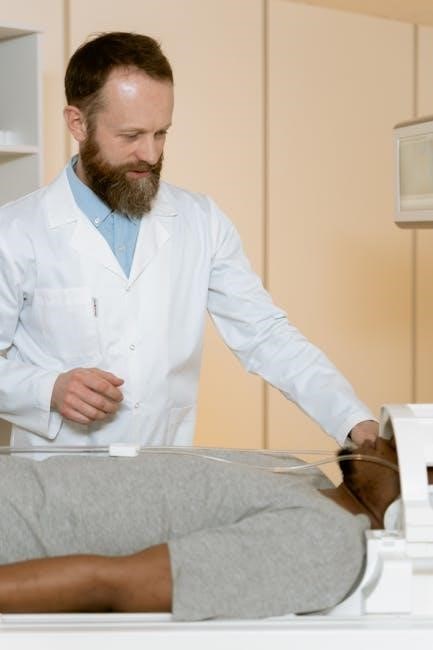
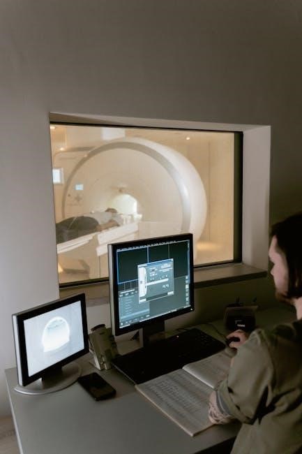
The manual integrates clinical examples and anatomical terminology to bridge the gap between lab work and real-world healthcare. Students learn to identify and describe structures using precise language, essential for medical communication. Why This Matters boxes explain how terms like “dorsal” or “ventral” apply to human anatomy and clinical diagnoses. For instance, understanding anatomical planes helps in interpreting MRI scans. This focus on terminology ensures students can discuss complex concepts accurately, preparing them for professional environments where clear communication is critical. The manual’s emphasis on clinical relevance makes anatomical learning practical and meaningful.
Additional Resources and Support
Pre-lab video coaching, detailed dissection videos, and Mastering A&P assignments provide comprehensive support, enhancing understanding and practical skills, ensuring a well-rounded educational experience.
Pre-Lab Video Coaching and Dissection Videos
The manual includes pre-lab video coaching to prepare students for lab activities and detailed dissection videos that guide through complex procedures. These resources provide visual and instructional support, helping students understand anatomical structures and techniques before engaging in hands-on dissections. The videos cover essential steps, anatomical terminology, and clinical relevance, ensuring a thorough understanding of each exercise. Additionally, bone, muscle, and organ dissection videos are integrated to enhance learning outcomes and reinforce key concepts. These digital tools, alongside Mastering A&P assignments, create a dynamic and interactive learning environment tailored to student success.
Interactive Features and Mastering A&P Assignments
The manual incorporates interactive features such as pre-lab video coaching, dissection videos, and bone, muscle, and organ identification activities. These tools enhance engagement and understanding of anatomical structures. Additionally, Mastering A&P assignments provide personalized learning experiences, reinforcing key concepts through interactive quizzes, labeling exercises, and clinical application questions. These digital resources complement traditional lab work, offering students a dynamic and immersive learning environment. By integrating these features, the manual ensures that students can apply theoretical knowledge to practical scenarios, fostering a deeper understanding of human anatomy and its real-world applications. This comprehensive approach supports academic success and professional development.
This manual effectively bridges theoretical knowledge with practical skills, providing a comprehensive understanding of human anatomy through cat dissections and fostering real-world applications for future professionals.
Importance of the Manual in Anatomy Education
The Human Anatomy Laboratory Manual with Cat Dissections is a cornerstone in anatomy education, offering a hands-on, visually engaging approach to learning complex anatomical structures. By using cats as models, students gain practical insights into human anatomy through detailed dissections and full-color illustrations. The manual’s clear, structured exercises and real-world applications make it an invaluable resource for both laboratory and independent study. Its accessibility and comprehensive coverage ensure that students develop a deep understanding of anatomical concepts, preparing them for future careers in healthcare and scientific fields. This manual is a essential tool for effective anatomy education.
Impact on Learning and Future Applications
The Human Anatomy Laboratory Manual with Cat Dissections significantly enhances learning by providing hands-on experience with anatomical structures. Students develop critical observational and tactile skills through dissections, which are vital for healthcare professionals. The manual’s focus on real-world applications bridges the gap between theoretical knowledge and practical scenarios, fostering critical thinking and problem-solving. By mastering anatomical terminology and concepts, students gain a strong foundation for careers in medicine, nursing, and research. This resource not only enriches academic understanding but also equips learners with skills essential for future professional success in anatomy-related fields.
Leave a Reply
You must be logged in to post a comment.